
X射线探测
X射线检测器中使用的闪烁体材料需要高X射线吸收效率和光输出,低余辉,透明性以及与光电转换器匹配的光谱响应。不同闪烁体的性能差异很大。为了与特定的CCD配合使用,我们应该综合考虑发光效率,光谱响应和其他特性,并选择合适的闪烁体材料。
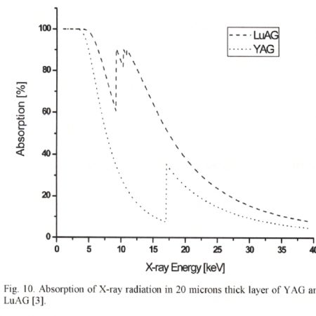
闪烁体中X射线辐射的平均吸收深度取决于光子能量和材料。材料透明度的优势随成像板的厚度而降低。如果闪烁体更薄,则平均吸收深度会更低,并且由于闪烁光子的横向扩展较少而导致的图像模糊较少,因此生成的图像会更清晰。因此,成像板越薄,图像中获得的分辨率越好。另一方面,随着闪烁体的厚度,检测效率降低。
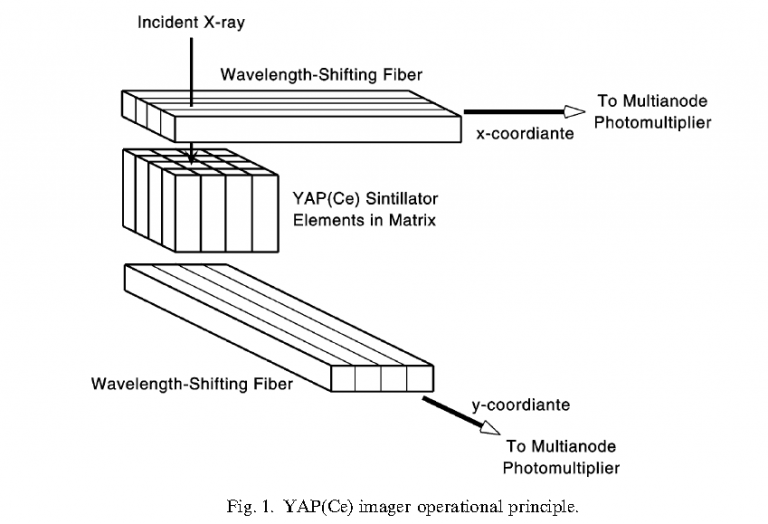
基于CCD的X射线探测器由闪烁体,光耦合器和CCD阵列组成。 它覆盖了CCD线性阵列上的闪烁体薄层。 闪烁体将X射线转换为对CCD敏感的可见光,然后将CCD转换为反映X射线在空间中分布的电信号。

应用于X射线探测的闪烁晶体
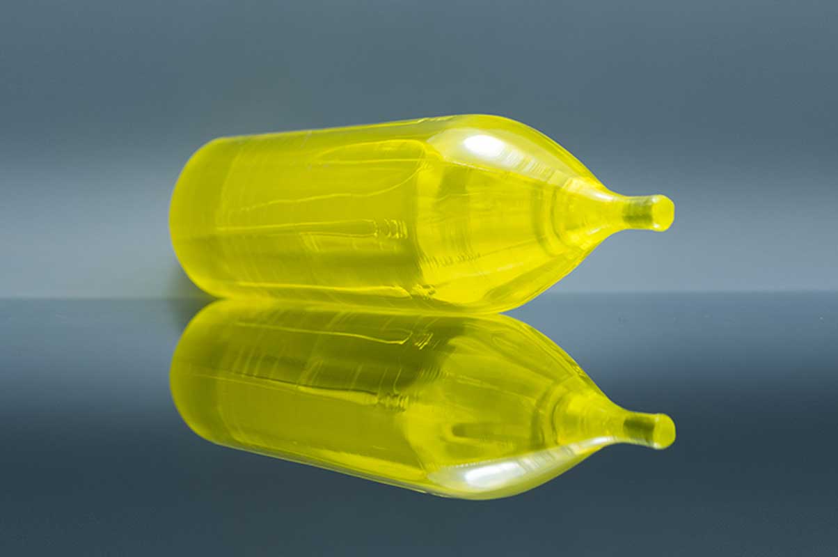
Ce:YAG
| 波长(最大发射)(nm) | 550 |
| 衰减时间(ns) | 70 |
| 发光量(光子/ keV) | 35 |
| 折光率 | 1.82@550nm |
| 辐射长度(cm) | 3.5 |
Ce:YAG是一种快速闪烁体,具有优异的机械和化学耐受性,其衰减时间约为70 ns,这使得高计数率成为可能,并且量子产率估计为 介于40至50 ph / keV之间,尽管光输出较小,但它的非吸湿性和较短的衰减时间与标准CsI:Tl闪烁体相比仍具有优势,最大发射波长(约550 nm)非常适合 PMT模块的光阴极的灵敏度。实验证明,YAG:Ce和LuAG:Ce屏幕适合于高空间分辨率的成像。所提出的成像系统的分辨率约为1微米。
参考文献
[1] An X-ray counting system based on YAG Ce scintillator
[2] Thin YAG:Ce and LuAG:Ce single crystal imaging plates used for high spatial resolution in X-ray imaging systems
[3] Similarity of trap state and thermoluminescence processes of Ce YAG for X-ray and UV irradiation
[4] High-resolution imaging of biological and other objects with an X-ray digital camera
[5] High-resolution application of YAG Ce and LuAG Ce imaging detectors with a CCD X-ray camera
Ce:YAP
| 波长(最大发射)(nm) | 370 |
| 衰减时间(ns) | 28 |
| 发光量(光子/ keV) | 25 |
| 相对于Nal(Tl)的光输出(%) | 60-70 |
| 折光率 | 1.95@370nm |
Ce:YAP晶体是出色闪烁体的特性包括:(i)30 ns的快速衰减时间,(ii)7.4 g / cm3的高密度,和(iii)约30%的高光产率 在NaI(Tl)闪烁体中 原铝酸钇(YAlO3:Ce or YAP:Ce)是快速发射闪烁体,主要用于PET和动物PET检测器中
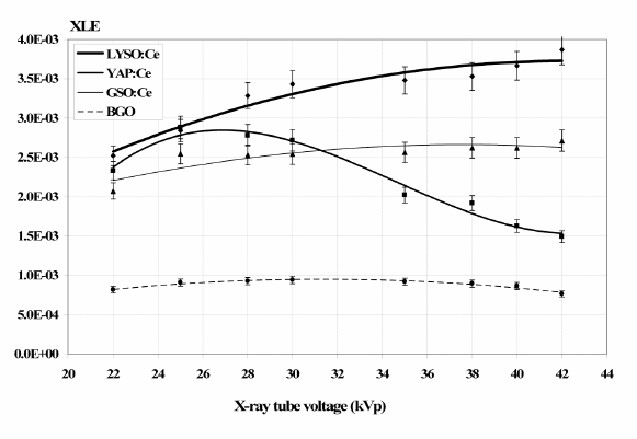
The x-ray luminescence efficiency (XLE) of Ce:LYSO, Ce:YAP, Ce:GSO and BGO as determined by the experimental data for x-ray tube voltages between 22–42kVp(mammography).Points: measured data, line: fitted curve.
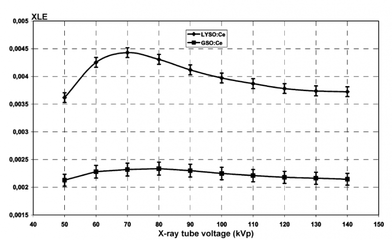
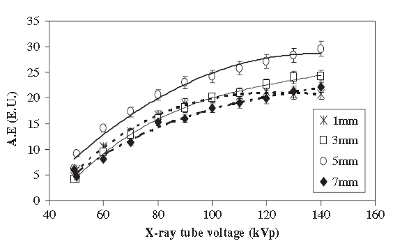
The x-ray luminescence efficieny (XLE) of Ce:LYSO, Ce:YAP, Ce:GSO and BGO as determined by the experimental data for x-ray tube voltages between 50–140 kVp (general radiography).Points: measured data, line: fitted curve.

参考文献
[1] A YAP(Ce) imager operated in high energy X-ray region
[2] Comparative evaluation of single crystal scintillators under x-ray imaging conditions
[3] Comparative Investigation of Ce Doped Scintillators in a Wide Range of Photon Energies Covering X-ray CT, Nuclear Medicine and Megavoltage Radiation Therapy Portal Imaging Applications
[4] ISPA tubes with scintillating YAP Ce windows X- and γ- ray imaging
[5] High-resolution application of YAG Ce and LuAG Ce imaging detectors with a CCD X-ray cameraMcps-range photon-counting X-ray computed tomography system utilizing an oscillating linear-YAP(Ce) photon detector
[6] Properties of a YAP Ce detector for high-energy X-ray counting experiments
Ce:LYSO

| 波长(最大发射)(nm) | 410 |
| 衰减时间(ns) | 40 |
| 发光量(光子/ keV) | 25 |
| 相对于Nal(Tl)的光输出(%) | 75 |
| 折光率 | 1.82@410nm |
基于镥-钇的闪烁体,例如LYSO:Ce,具有很高的有效原子序数,是非吸湿的,快速发射的材料,并且有望用于正电子发射成像仪。
Ce: LYSO是混合的LSO / YSO(5-10%)非吸湿性晶体,具有高密度(7.1 g / cm3),高光输出(30000 ph / MeV),良好的能量分辨率(10%)和短衰减 时间(40 ns)。 尽管Ce: LYSO和Ce: LSO表现出相似的行为,但就衰减方案而言,在低能(35 kV)X射线激发下,Ce: LYSO的光产率似乎比LSO高约20%。

参考文献
[1] A comparative study of the luminescence properties of LYSO Ce, LSO Ce, GSO Ce and BGO single crystal scintillators for use in medical X-ray imaging
[2] Comparative evaluation of single crystal scintillators under x-ray imaging conditions
[3] Comparative Investigation of Ce Doped Scintillators in a Wide Range of Photon Energies Covering X-ray CT, Nuclear Medicine and Megavoltage Radiation Therapy Portal Imaging Applications
[4] Evaluation of the light emission efficiency of LYSO Ce scintillator under X-ray excitation for possible applications in medical imaging
[5] Improving Ce3+ doped scintillating materials for medical imaging applications
[6] Investigation of luminescence emission properties of LYSO Ce and LuYAP Ce single crystal scintillators under x-ray exposure for use in medical imaging
[7] Luminescence Properties of LYSO ce and GSO Ce Single Crystal Scintillators Under X-Ray Excitation for Use in Medical Imaging Systems
[8] Scintillator efficiency study with MeV x-rays
Ce:GAGG
| 波长(最大发射)(nm) | 520 |
| 波长范围(nm) | 475-800 |
| 衰减时间(ns) | 90 |
| 发光量(光子/ keV) | 50 |
| 折光率 | 1.95@540nm |
Ce:GAGG (Ce:Gd3Al2Ga3O12) 闪烁晶体,具有高密度(6.69 g/cm-3),快速闪烁响应(〜90 ns)和高光产率(〜46000 ph / MeV) )。 与目前许多高效的闪烁体相比,GAGG:Ce具有非吸湿性(如LaBr3:Ce,LaCl3:Ce,CsI和NaI:Tl晶体),并且不具有天然放射性(如基于based的材料)。 GAGG:Ce发出520nm的光子,有效原子序数等于54.4。

参考文献
[1] X-ray Luminescence Efficiency of GAGG Ce Single Crystal Scintillators for use in Tomographic Medical Imaging System
[2] Light Emission Efficiency of GAGG Ce Single Crystal Under X-ray Radiographic Conditions
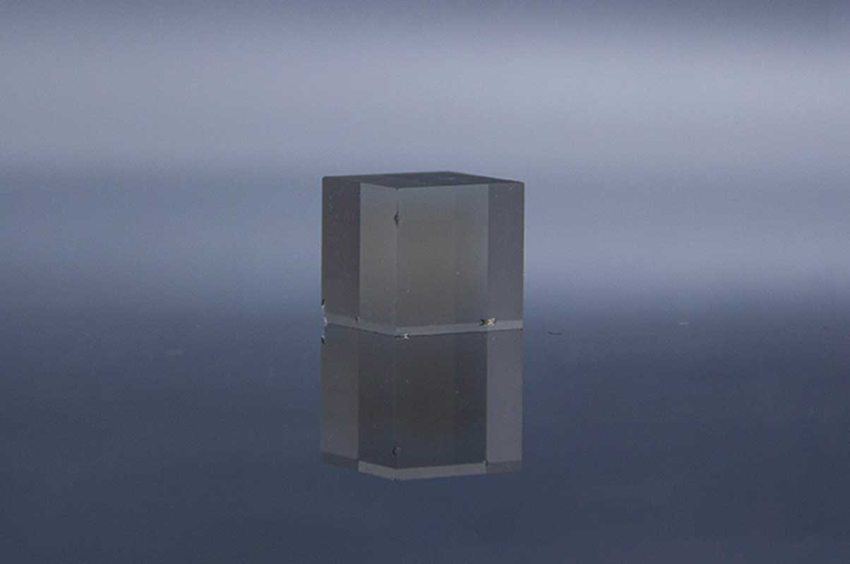
Ce:LuAG
| 波长(最大发射)(nm) | 535 |
| 波长范围(nm) | 475-800 |
| 衰减时间(ns) | 70 |
| 发光量(光子/ keV) | 25 |
| 折光率 | 1.84@633nm |
高分辨率成像系统是由高灵敏度数字CCD摄像机和光学系统与薄闪烁成像屏组成。屏幕可以包括LuAG:Ce无机闪烁体。
[参考文献1]比较了两种屏幕的发光效率(每keV可见光子数)和空间分辨率。在第一个实验中,CCD检测到的光在CCD中心200μ200像素的平方ROI中平均。这个Ce:YAG发射值17507(每秒CCD像素中的电子数)和Ce:LuAG给出值26452,大约是Ce:YAG。这个Ce:LuAG单曲晶体比钇铝石榴石(密度:6.73–4.57 g/cm3)并且X射线被LuAG吸收得更强(在1到40 keV范围内吸收的X射线辐射(光子)是X射线辐射衰减系数的1.7倍)。这个Ce:LuAG屏幕具有比Ce:YAG屏使图像的信噪比更好地应用于成像系统中。
[参考文献2]使用Ce:LuAG 20 mm屏幕拍摄图像。CCD相机的有效像素尺寸为0.74mm。X射线微聚焦源的工作电压为40kV / 2mA。图像采集时间为5s,对25个图像样本进行平均。在从源发出的光子通量转换后,Ce:LuAG产生的光的测量强度约为Ce:YAG值的1.51倍。用CCD检测光,并在200×200像素的平方ROI中求平均值。与Ce:YAG(密度:6.73–4.57g / cm3)相比,Ce:LuAG单晶密度更高,LuAG对X射线的吸收更强(X射线的平均吸收量是1.7倍)。1至40keV之间的范围)
参考文献
[1] High-resolution application of YAG Ce and LuAG Ce imaging detectors with a CCD X-ray camera
[2] High-resolution imaging of biological and other objects with an X-ray digital camera
[3] Thin YAG Ce and LuAG Ce single crystal imaging plates used for high spatial resolution in X-ray imaging systems
Tl:CsI
| 波长(最大发射)(nm) | 410 |
| 衰减时间(ns) | 40 |
| 发光量(光子/ keV) | 25 |
| 相对于Nal(Tl)的光输出(%) | 75 |
| 折光率 | 1.82@410nm |
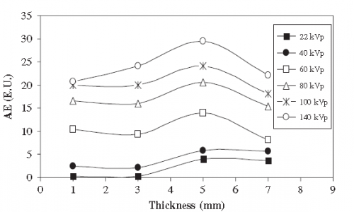
CsI(Tl)的最重要特征是其高光产率(≥104光子/ MeV),并且其发射光谱在约550 nm处具有最大值,与非晶和结晶硅光电二极管完全兼容。研究了CsI:Tl单晶闪烁体的发光效率与X射线乳腺摄影和一般X射线成像所用能量范围内的晶体厚度和X射线管电压的关系。
[参考文献1]绝对发光效率,这是在X射线成像(40–140 kV)和乳房X线照片成像(22–49 kV)中使用的各种X射线能量中通过实验确定的。使用以下设备通过X射线对晶体进行辐照:(i)具有钨阳极靶和2mm Al滤光片的飞利浦X射线装置和(ii)带有钼阳极靶和钼滤光片的通用电子Senographer DMR X射线乳腺摄影装置,使用前面描述的实验装置,对晶体进行X射线辐照。
[参考文献2]为了满足这些需求,我们使用结构化的CsI(TI)闪烁体与快速帧1K x 1K CCD耦合,开发了一种原型快速X射线成像系统。该系统已成功用于以12位动态范围以每秒1000帧(fps)的速率捕获1024 x 64像素x射线图像。该系统超越了通常以30 fps的速率运行的当前高速X射线成像系统的功能。CsI(TI)(59,000个光子I MeV)的高x射线转换效率使这些屏幕成为当前x射线衍射应用的一个极好的选择,在x射线衍射中,某些重要的衍射峰往往很弱,需要具有高信噪比的成像屏幕才能正确识别。

参考文献
[1] A systematic study of the performance of the CsI Tl single-crystal scintillator under X-ray excitation
[2] high-speed-xray-imaging-camera-using-structured-csitl-scintillator
[3] Scintillator efficiency study with MeV x-rays
[4] Structured CsI(Tl) scintillators for X-ray imaging applications
[5] Validation of columnar CsI x-ray detector responses obtained with hybridr , a CPU-GPU Monte Carlo code for coupled x-ray, electron, and optical transport
CdWO4
| 波长(最大发射)(nm) | 490 |
| 衰减时间(ns) | 14000 |
| 发光量(光子/ keV) | 12-15 |
| 相对于Nal(Tl)的光输出(%) | 50 |
| 折光率 | 2.2-2.3 |
钨酸镉(CdWO4,CWO)晶体是此类应用的候选者之一,因为它具有较高的有效Z值,高密度,比BGO更高的光产率以及非常低的余辉。CWO晶体的辐射硬度高达105 rad。由于这种优势,CWO闪烁体被广泛用于X射线CT中。
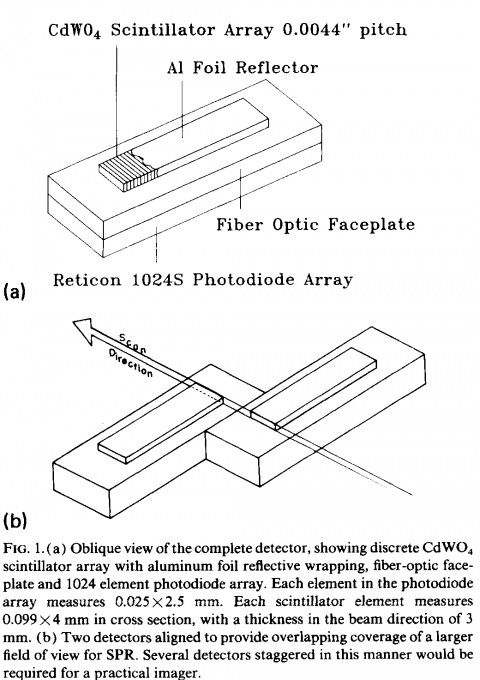
[参考文献1]描述了一种用于X射线成像的高分辨率X射线检测器阵列,它由CdWO4闪烁体的离散阵列和带有光纤面板的Reticon 1024S光电二极管阵列组成。它开发了一种非常高分辨率的离散闪烁体阵列,用于扇形束X射线成像系统。
参考文献
[1] High-resolution digital x-ray detector utilizing a discrete array of CdWO4 scintillators and a self-scanned photodiode array
[2] Large Size CdWO4 Crystal for Energetic X- and γ Ray Detection
[3] X-RAY STUDY OF CHARACTERIZATION OF THE PbWO4 AND CdWO4 SINGLE CRYSTALS
GOS
| 波长(最大发射)(nm) | 510 |
| 衰减时间(us) | 5.5 |
| 相对光输出(%) | 80 |
| 余辉(%) | <0.01 |
| 发光强度(keV) | 27.5 |

| GOS(Gd2O2S) | Density (g/cm3) | Wavelength of max emission (nm) | Decay constant (ns) |
| Ref[1] | 7.34 | 510 | 5.5μs |
参考文献